Functional Spatial Analysis for risk assessment in IPMN published in Cancer Informatics

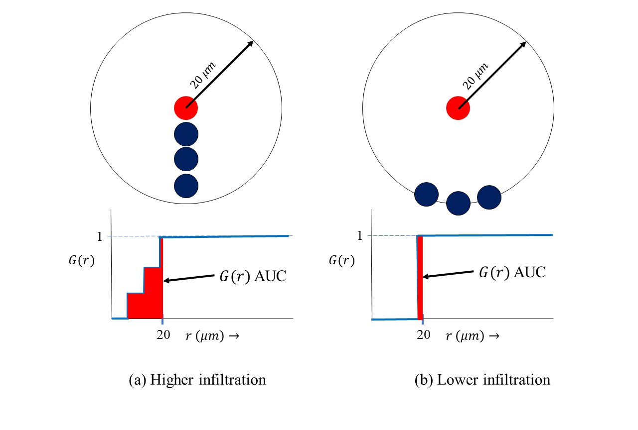
Our paper titled “A Functional Spatial Analysis Platform for Discovery of Immunological Interactions Predictive of Low-Grade to High-Grade Transition of Pancreatic Intraductal Papillary Mucinous Neoplasms” has been published in Cancer Informatics’ Special issue on Ensemble Learning and Deep Learning in Cancer Genomic and Imaging Data. In this work, we enhance our G-function based spatial analysis framework by incorporating ideas from functional data analysis, to build a tool more effective at representing the structure in the G-function. We show that spatial metrics derived from our functional spatial platform is able to predict the risk of progression in IPMN’s with a higher accuracy than simpler metrics such as counts or the G-function AUC alone. AN ensemble of models built using counts and the proposed G-function MFPCA metric performs the best. The paper can be found here.


Spatial tumor:Treg interaction predicts poor survival in Non-Small Cell Lung Cancer accepted to Lung Cancer journal

Our paper on using the spatial G-function to quantify tumor:immune interactions in multiplexed Immunofluorescence images from Non-Small Cell Lung Cancer patients has been accepted to Lung Cancer! [Paper link] We made two contributions in this paper: 1) Repurposed the G-function used in ecology to study predator-prey relationships for studying tumor-immune cell interactions in NSCLC, and 2) That a simple area under the curve (AUC) metric derived from the G-function representing tumor cell- regulatory T cell interaction correlates with poor outcome for patients, independently of clinical variables that doctors currently use! (such as age, smoking history, number of positive lymph nodes, size of tumor etc.)

Since the G-function is a unique signature of immune infiltration, my hope is that doctors will now incorporate our fast and easy-to-use algorithm in clinical practice!

Spatial co-localization work in Merkel Cell Carcinoma accepted to USCAP 2018

Our work with Dr. Phyu Aung, Dr. Michael Tetzlaff (Dept. of Pathology, M.D Anderson Cancer Center) and Dr. Ignacio Wistuba’s lab (Dept. of Translational Molecular Pathology) on studying the co-localization of B7-H3 expression in endothelial cells of Merkel Cell Carcinoma (MCC) patients, has been accepted as an abstract to the prestigious United States and Canadian Academy of Pathology (USCAP) Annual Meeting in March 2018. MCC is a very aggressive form of skin cancer, and patient response is closely linked to immune system integrity. The B7-H3 biomarker has been shown to be a potent inhibitor of the human body’s immune response. We used our novel spatial infiltration metric on multiplexed Immunofluorescence images of resected MCC tumors, to compute the extent of B7-H3 expression in endothelial cells. This quantification enables us to now directly study the impact of B7-H3 colocalization on patient outcome, potentially allowing us to design optimal immunotherapy regimens for individual patients with MCC!!

Radiomics based ORN prediction work accepted in ASTRO-HNC 2018

Our work with Dr. Hesham Elhalawani (Dept. of Radiation Oncology, M.D Anderson Cancer Center) on early prediction of Osteo-radio-necrosis (ORN) using radiomics has been accepted to the American Society for Radiation Oncology’s Head and Neck cancer (ASTRO-HNC) symposium in 2018. We show that a functional principal component analysis (FPCA) of radiomic features extracted at multiple time points before and after radiotherapy, can predict for ORN development in patients. We significantly outperform prediction models based on pre-radiotherapy images and delta radiomics (current practice). We envisage our FPCA screening tool can be used by radiologists to optimize radiation plan for patients undergoing radiotherapy!